 how-to
Why pH Balance Affects Peptide Stability: A Lab Guide
Table of Contents
- Why pH Balance Affects Peptide Stability
- How pH Affects Peptide Structure and Charge State
- Peptide Degradation at High pH: Hydrolysis and Side-Chain Changes
- Acidic vs. Alkaline Conditions: Which Is Worse for Your Peptide?
- Peptide Reconstitution pH: Solvent Choice and Water Balance
- Peptide Storage and Stability: Temperature, Light, Oxygen, and Container Materials
- Step-by-Step pH Troubleshooting Workflow for the Lab
- Conclusion: Prioritizing pH Control for Reproducible Results
- Frequently Asked Questions
Last Updated: October 7, 2026
Why pH Balance Affects Peptide Stability
pH balance is the most overlooked factor in peptide handling. Get it wrong, and your peptide can degrade before your assay begins.
Peptides are chains of amino acids carrying electric charges that shift with pH, altering charge state, shape, and stability.
Peptide stability is the ability of a peptide to keep its molecular structure and activity under given storage and handling conditions. pH is one of the biggest levers on that stability, often more important than temperature alone.
How pH Affects Peptide Structure and Charge State
pH sets the charge on every ionizable group along the peptide backbone and side chains, deciding how it folds, dissolves, and breaks down.
Each side chain has its own pKa, the pH where it flips between charged and neutral. Near those values, small shifts flip charges on and off, making solubility and structure a moving target.
Ionization, Isoelectric Point, and Molecular Structure
The isoelectric point (pI) is the pH where a peptide carries no net charge. Solubility often bottoms out there because no charge repulsion keeps molecules apart.
Away from the pI, charges keep molecules dissolved; near it, they clump. That is why the same peptide can dissolve cleanly at one pH and turn cloudy at another.
- Low pH: acidic groups go neutral, basic groups go positive
- High pH: basic groups go neutral, acidic groups go negative
- At the pI: net charge is zero, and solubility tends to bottom out
Peptide Degradation at High pH: Hydrolysis and Side-Chain Changes
High pH accelerates peptide breakdown, mainly through hydrolysis of peptide bonds: hydroxide attacks the backbone and the chain can split.
The peptide backbone is the repeating N-C-C pattern holding the chain together. When a peptide bond breaks, you lose the intact molecule and gain fragments, lowering the effective concentration of what you intended to study.
Epimerization and Other Chemical Changes
Epimerization converts an amino acid from one stereochemical form to another at a single chiral center. Extreme pH speeds this up, and it is hard to detect without the right analysis.
Other side-chain changes matter too: basic residues can react under alkaline conditions, and some side chains oxidize when pH and oxygen combine. These alter the molecule without breaking the chain.
- Hydrolysis: splits the peptide bond, shortens the chain
- Epimerization: flips stereochemistry, changes activity
- Oxidation: modifies side chains, especially sulfur-containing ones
Acidic vs. Alkaline Conditions: Which Is Worse for Your Peptide?
Neither extreme is safe, but they damage peptides differently. Alkaline conditions drive hydrolysis and epimerization faster; acidic conditions are gentler on the backbone but can still cause problems.
Most peptides tolerate mild acidity better than strong alkalinity, which is why dilute acetic acid is such a common reconstitution solvent: it keeps the peptide charged and dissolved without pushing the backbone toward hydrolysis.
But "acidic is safer" is not a blanket rule, some sequences are acid-sensitive, some base-sensitive. The right answer depends on the specific residues in your peptide.
| Condition | Main Risk | Typical Effect |
|---|---|---|
| Strongly acidic | Side-chain changes, cleavage at sensitive sites | Usually milder on backbone |
| Mildly acidic | Low, if matched to peptide | Best for many peptides |
| Neutral | Mixed, depends on pI | Can trigger aggregation near pI |
| Mildly alkaline | Hydrolysis, deamidation | Gradual loss of intact peptide |
| Strongly alkaline | Fast hydrolysis, epimerization | Rapid degradation |
Peptide Reconstitution pH: Solvent Choice and Water Balance
The solvent you reconstitute with sets the starting pH and follows the peptide through every step.
Reconstitution is dissolving a lyophilized peptide powder in liquid to create a working solution. The solvent decides the pH, ionic strength, and how stable the peptide stays.
Pure water has essentially no buffer capacity, so a trace of residual acid or base can swing pH by a full unit or more.
Acetic Acid, Bacteriostatic Water, and Buffer Capacity
Dilute acetic acid keeps many peptides charged and dissolved.
Bacteriostatic water contains 0.9% benzyl alcohol to limit microbial growth, useful for multi-use vials. But benzyl alcohol can oxidize methionine and cysteine residues and interfere with cell-based assays, so check compatibility before committing a full vial.
Buffer capacity is a solution's ability to resist pH change when acid or base is added, keeping pH stable across handling, dilution, and storage. Common choices include:
- Sodium acetate (pKa ~4.76): good for acidic working ranges, compatible with many reverse-phase HPLC methods
- Sodium phosphate (pKa ~7.2): neutral-range workhorse, but can precipitate in high-organic HPLC mobile phases
- Tris (pKa ~8.06): alkaline range, but the primary amine can react with some reagents and its pKa drifts strongly with temperature
A practical rule: pick a buffer whose pKa sits within one pH unit of your target. Outside that window, buffer capacity collapses and pH drifts with every addition.
- Match solvent pH to keep the peptide away from its pI
- Use dilute acetic acid (0.1% to 1%) for peptides that dissolve poorly near neutral pH
- Choose bacteriostatic water only when preservative compatibility is confirmed
Measuring pH Accurately at Small Volumes
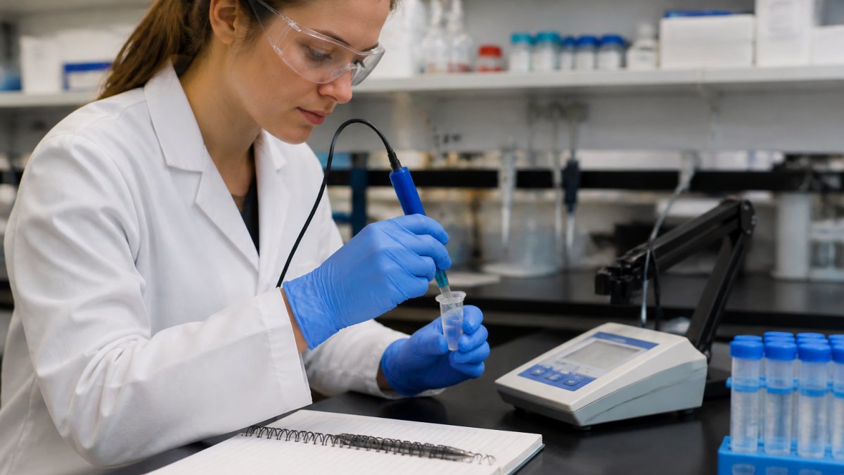
Standard glass pH electrodes need 3 to 5 mL of sample, far more than a typical reconstituted peptide vial holds.
Options that work at small volumes:
- Micro pH electrodes: bulb diameters of 1 to 3 mm, usable with 50 to 200 microliters in a microcentrifuge tube. Require two- or three-point calibration with fresh standards each session.
- Flat-surface or ISFET probes: read from a 20 to 50 microliter drop placed on the sensor. Fast response, but drift more than glass electrodes and need frequent recalibration.
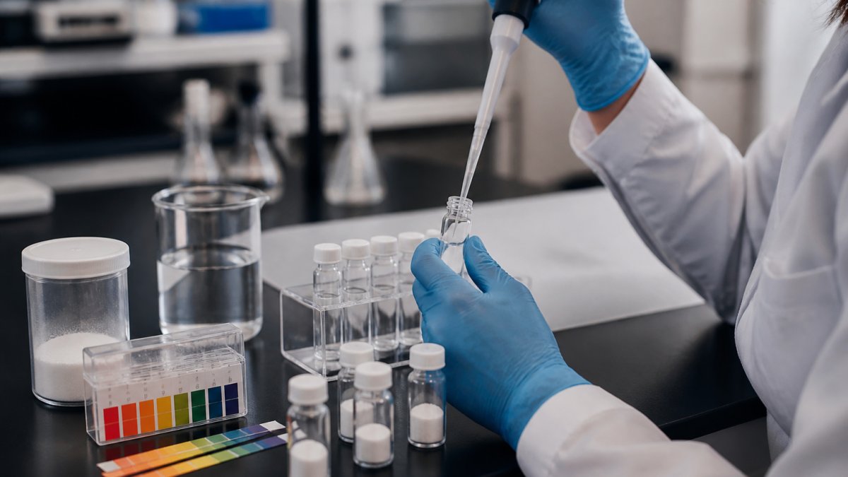
- pH indicator strips: only useful for coarse confirmation (roughly 0.5 pH unit resolution). Not adequate for stability-critical work.
Calibration discipline matters more than the meter. Use fresh pH 4.00, 7.00, and 10.01 standards, calibrate at your measurement temperature, and re-check the slope.
Measure pH after reconstitution and again after any dilution or buffer exchange. Dilution with unbuffered water can shift pH by a full unit even when the stock was correct.
Peptide Storage and Stability: Temperature, Light, Oxygen, and Container Materials
pH is not the only variable. Temperature, light, oxygen, and the container all shape how long a peptide stays intact.
Lower temperatures slow chemical reactions, so cold storage slows degradation. But cold storage does not fix a bad pH: a peptide at the wrong pH still degrades, just more slowly.
Light and oxygen drive oxidation, especially for peptides with sulfur-containing residues; amber vials and inert headspace help.
- Temperature: store cold to slow reaction rates
- Light: use amber or foil-wrapped vials
- Oxygen: limit headspace and consider inert gas
For documented integrity, lot-specific records and third-party testing give you a paper trail. Bluegrass Research supplies research-grade peptides with lot-specific COA documentation, third-party analytical testing, and transparent verification records so you can trace purity and handling for each batch.
Step-by-Step pH Troubleshooting Workflow for the Lab
When a peptide will not dissolve, turns cloudy, or loses activity, work through pH systematically instead of guessing. This workflow adds decision criteria and analytical confirmation steps most guides skip.
Step 1: Calculate or Look Up the pI
Use the peptide sequence and a pI calculator (ExPASy, IPC, or equivalent) to estimate the isoelectric point. Calculated pI values assume ideal behavior and can be off by 0.5 to 1.0 pH unit for peptides with unusual residues or post-translational modifications, so treat the number as a starting estimate, not a fact.
Step 2: Measure the Actual pH
Use a micro electrode calibrated the same day with fresh pH 4.00, 7.00, and 10.01 standards. Record the temperature. Do not assume the pH matches the label or the solvent's nominal value.
Step 3: Compare Working pH to pI
If the working pH sits within roughly 1 pH unit of the pI, solubility problems are likely. Move the pH at least 1.5 to 2 units away from the pI in the direction that keeps the peptide charged.
Step 4: Adjust in Small Steps
Add dilute acid (0.1% acetic acid or 0.1 M HCl) or dilute base (0.1 M NaOH or ammonium hydroxide) in 1 to 2 microliter increments to a small aliquot, re-measuring after each addition. Never adjust the whole batch until you confirm the target pH on a test aliquot.
Step 5: Observe the Response
- Clears as pH moves away from pI: solubility problem, not degradation. Scale the adjustment to the full batch.
- Clears with gentle warming (25 to 37 degrees C): likely reversible aggregation. Cool back down and confirm it stays clear.
- Stays cloudy at every pH: likely irreversible aggregation or chemical degradation. Proceed to analytical confirmation.
Step 6: Confirm with Analytical Methods
Visual clarity is not evidence of integrity. If the solution stays cloudy, activity drops, or you need documented proof, run one or more of these:
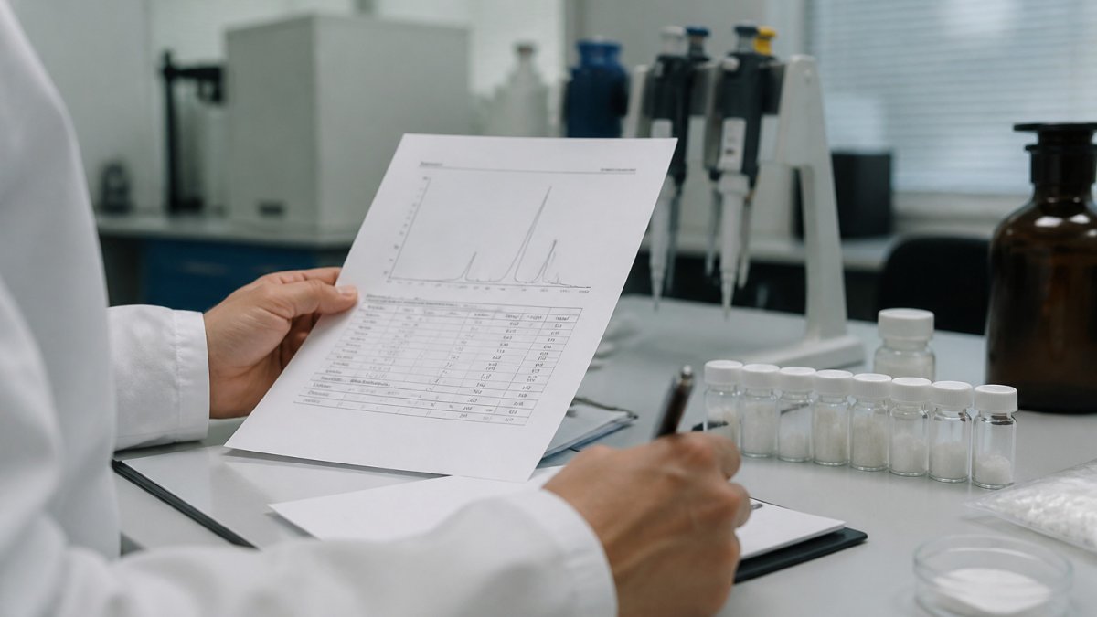
- Reverse-phase HPLC: separates intact peptide from fragments and detects hydrolysis products. A shift in retention time or the appearance of new peaks signals degradation.
- LC-MS or MALDI-TOF: confirms molecular weight. A mass shift of +18 Da suggests hydrolysis; +1 Da suggests deamidation; +16 Da suggests oxidation.
- Circular dichroism (CD): detects secondary structure changes that do not alter mass.
Distinguishing Reversible Aggregation from Irreversible Degradation
Aggregation is the clumping of peptide molecules, and it can be reversible or not. Reversible aggregation often clears when you change pH, temperature, or concentration.
Observable signs that point one way or the other:
| Observation | Likely Reversible Aggregation | Likely Irreversible Degradation |
|---|---|---|
| Clears with pH shift | Yes | No |
| Clears with gentle warming | Often | Rarely |
| Clears with dilution | Often | No |
| HPLC shows single peak | Yes | No |
| Mass spec matches expected mass | Yes | No |
| Activity restored after clarification | Yes | No |
The practical test is simple. If a pH shift or gentle warming restores a clear solution and full activity, you had aggregation. If nothing restores it, you likely have hydrolysis, epimerization, or another chemical change.
A Note on Safety and Documentation
Handle dilute acids and bases with appropriate PPE, and neutralize waste before disposal per your institution's chemical hygiene plan. Log every pH measurement, adjustment, and analytical result against the lot number to trace any stability issue back to its source.
Conclusion: Prioritizing pH Control for Reproducible Results
pH control is not a minor step. It decides whether your peptide dissolves, stays intact, and gives you trustworthy data.
The workflow is straightforward: know the pI, measure the real pH, adjust in small steps, and confirm with analysis when something looks wrong.
When you need verified starting material, Bluegrass Research supplies research-grade peptides with lot-specific COA documentation, third-party analytical testing, and discreet shipping with no external branding.
Shop Now
Frequently Asked Questions
How does pH affect peptide structure?
pH determines the electric charge on a peptide's side chains and backbone. At a given pH, each ionizable group is either protonated or deprotonated, which changes the peptide's overall charge state and molecular structure. Near the isoelectric point, the net charge is zero, reducing solubility and promoting aggregation. Extreme pH can also disrupt hydrogen bonding and lead to hydrolysis of peptide bonds or side-chain changes. Because charge state influences both structural integrity and solubility, pH is a primary variable in peptide stability.
What disrupts peptide bonds?
Peptide bonds are disrupted by hydrolysis, a reaction that splits the bond between amino acids. Hydrolysis is accelerated by extreme pH (especially high pH), elevated temperature, and certain enzymes. In acidic conditions, hydrolysis can also occur, particularly at aspartic acid residues. To protect peptide bonds, keep the pH near the peptide's optimal stability range, store lyophilized powders cold and dry, and avoid prolonged exposure to harsh conditions. Using a buffer with adequate capacity helps maintain a stable pH.
Does reconstitution solvent affect peptide stability?
Yes. The reconstitution solvent sets the initial pH and can influence solubility, aggregation, and degradation rate. Acetic acid is often used for basic peptides, while ammonium hydroxide may be needed for acidic ones. Bacteriostatic water provides a preservative but can affect pH slightly. Always check the peptide's solubility instructions and, when possible, measure the pH of the final solution. If the pH is outside the stable range, adjust with small amounts of acid or base, or choose a different buffer.
How can researchers monitor pH-related changes in peptide stability?
Monitor stability by measuring pH at small volumes with a microelectrode, then tracking changes over time using analytical methods. Reverse-phase HPLC can detect hydrolysis and side-chain modifications, while size-exclusion chromatography or dynamic light scattering can reveal aggregation. Mass spectrometry confirms molecular weight changes from degradation. For a practical workflow, prepare samples at different pH values, incubate under controlled conditions, and analyze at set intervals. Document everything, including buffer composition and temperature, to correlate pH with stability outcomes.