 how-to
How to Troubleshoot Peptide Precipitation in Buffer
Table of Contents
- Peptide Precipitation: Why It Happens During Buffer Prep
- Step 1: Diagnose the Cause Before You Adjust Anything
- Step 2: Optimize pH and Ionic Strength for Peptide Solubility
- Step 3: Choose the Right Buffer Additives to Prevent Peptide Aggregation
- Step 4: Master Buffer Exchange Techniques Without Losing Your Peptide
- Bacteriostatic Water vs. Sterile Water for Peptides: What Changes in Buffer Prep
- Step 5: Recovery Protocols When Precipitation Has Already Occurred
- Common Mistakes That Trigger Peptide Precipitation
- Frequently Asked Questions
Last Updated: October 2, 2026
Peptide Precipitation: Why It Happens During Buffer Prep
The problem usually starts before the peptide touches the buffer. Most researchers blame the compound, but the buffer recipe is the culprit far more often.
Most guides tell you to add more solvent, which hides the real cause; the peptide crashes out again at the next step.
Below, we'll show you how to troubleshoot peptide precipitation during buffer preparation, from the first sign of cloudiness to full recovery: diagnose, adjust, stabilize, exchange, recover.
The Difference Between Precipitation and Aggregation
Precipitation is peptide leaving solution and forming a solid you can see or spin down. Aggregation is peptide molecules sticking to each other while still in solution, often invisible until the clumps grow large enough to fall out. The two travel together: a peptide that aggregates will usually precipitate once the clumps get big enough.
Peptide-Specific vs. Protein-Specific Behavior
Short peptides behave differently from large proteins, and the standard protein playbook does not always transfer.
- Size: Peptides are small, diffuse fast, and respond quickly to pH shifts.
- Structure: Most short peptides lack a folded core, so they are less prone to classic denaturation.
- Hydrophobicity: A single hydrophobic stretch can drive the whole molecule out of solution.
- Charge: The isoelectric point matters more for peptides because there is less surface area to buffer the effect.
That last point trips people up. A protein might tolerate a pH near its isoelectric point. A short peptide at the same pH can drop out of solution within minutes.
Step 1: Diagnose the Cause Before You Adjust Anything
Stop and look before you change the buffer. Random fixes make the problem harder to trace.
Work through this checklist in order:
- Note the pH of the current buffer.
- Note the salt concentration and buffer species.
- Check the temperature of the solution and the room.
- Look at how the peptide was added (dumped in, or slowly).
- Check whether the peptide was fully dissolved before mixing.
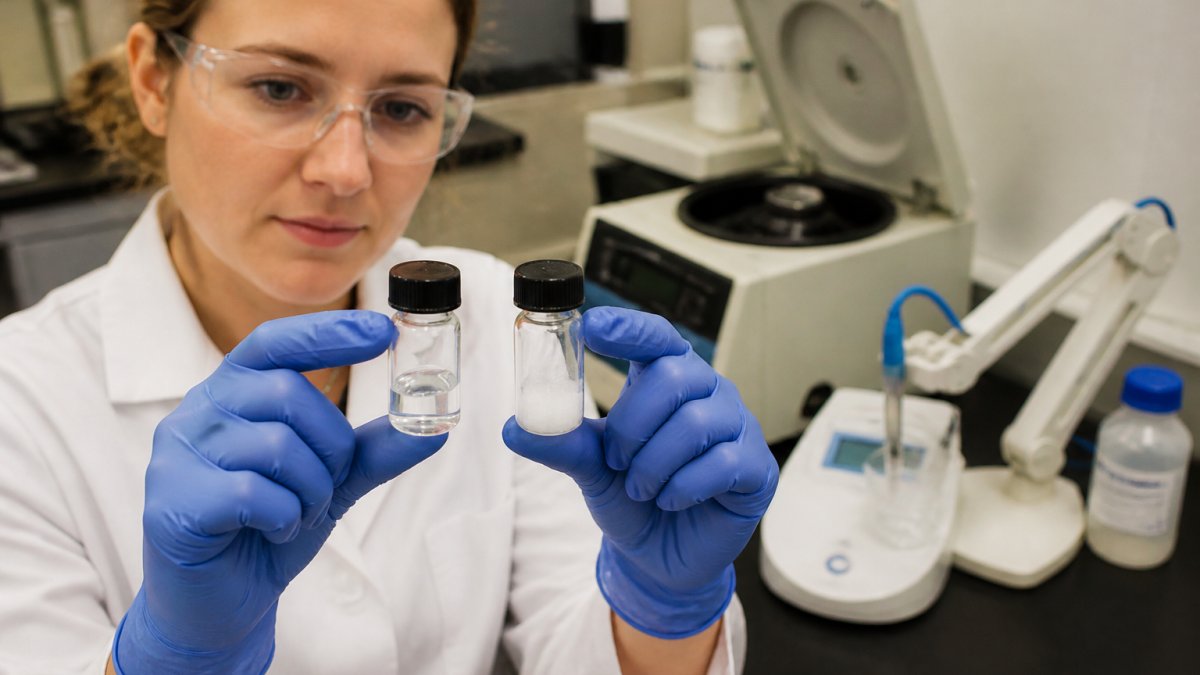
Visual Identification Guide: Turbidity, Clumping, and Pellet Formation
What you see tells you what went wrong.
| What You See | Likely Cause | First Fix |
|---|---|---|
| Hazy, uniform turbidity | pH near isoelectric point | Shift pH by 1-2 units |
| Stringy clumps | Hydrophobic aggregation | Add arginine or a mild detergent |
| Fine white pellet after spin | True precipitation | Redissolve at higher pH or add solvent |
| Cloudiness that clears on warming | Temperature-driven | Keep buffer at room temp |
Turbidity usually points to a pH problem, clumping to hydrophobicity, and a clean pellet to a solubility limit a pH shift can often reverse.
Step 2: Optimize pH and Ionic Strength for Peptide Solubility
Peptide solubility optimization starts with pH. Move the buffer pH at least one unit away from the peptide's isoelectric point, where the peptide carries no net charge, so molecules attract instead of repel and clump. Ionic strength matters just as much: low salt lets peptides stick to each other, while too much salt can force them out, an effect called salting out.
A practical starting range:
- pH: 1-2 units away from the isoelectric point
- Salt: 50-150 mM sodium chloride as a starting point
- Buffer species: match the pKa to your target pH
Test small aliquots before committing the whole batch. A 1 mL test tube tells you more than a full-scale prep gone wrong.
Step 3: Choose the Right Buffer Additives to Prevent Peptide Aggregation
Buffer additives to prevent peptide aggregation interrupt the interactions that pull peptides together, hydrophobic contacts, electrostatic bridging, and surface adsorption. Add them one at a time and titrate on a small aliquot before committing the batch.
Working Concentration Ranges
These are common starting ranges from peptide and protein formulation work. Treat them as starting points, not targets, and confirm compatibility with your downstream assay.
| Additive | Typical Working Range | Mechanism | Watch-Outs |
|---|---|---|---|
| L-Arginine (HCl or free base) | 50-500 mM | Suppresses hydrophobic and electrostatic self-association; weakens protein-protein contacts | Can raise buffer conductivity and interfere with ion-exchange; free base shifts pH |
| Glycerol | 5-20% v/v | Preferential hydration; raises the free energy of unfolding | Raises viscosity, slows filtration, can interfere with CD and some mass spec workflows |
| Sucrose or trehalose | 5-10% w/v | Preferential exclusion; stabilizes folded state during freeze-thaw | Adds osmolality; can crystallize at cold temperatures |
| Polysorbate 20 or 80 | 0.01-0.05% w/v | Competes with peptide for hydrophobic surfaces; reduces interfacial aggregation | Peroxide content in polysorbates can oxidize methionine and tryptophan residues |
| Urea or guanidine HCl | 1-4 M (denaturant) | Solubilizes aggregated material by disrupting H-bonds | Denatures the peptide; only useful if you plan to refold |
Peptide-Specific Notes
Short peptides respond differently than large proteins to the same excipient. Key patterns:
- Arginine is the first-line choice for peptides. It is small, charged, and does not require a folded structure to work. For hydrophobic peptides, 100-300 mM arginine often holds material in solution where a protein would need a detergent.
- Detergents help less than you would expect. Peptides lack the large hydrophobic surface patches detergents shield. Use polysorbate only when surface adsorption (to tubing or filter membranes) is the suspected cause.
- Sugars protect against freeze-thaw, not pH-driven precipitation. If your peptide crashes when the buffer pH is wrong, fix the pH first.
- Glycerol is a poor fit for hydrophobic peptides. It can reduce solubility of very nonpolar sequences by lowering the solution's dielectric constant.
Order of Addition
Add excipients to the buffer before the peptide, not after. Pre-mixed buffer gives the peptide a friendly environment from first contact. If you must add an excipient to an already-dissolved peptide, do it dropwise with gentle mixing and watch for turbidity.
Step 4: Master Buffer Exchange Techniques Without Losing Your Peptide
Buffer exchange is where most peptide is lost. Dialysis, ultrafiltration, and centrifugation-based exchange each have distinct failure modes; the right choice depends on peptide size, concentration, and tolerable dilution.
Match the Method to the Peptide
| Method | Best For | Typical MWCO | Key Failure Mode |
|---|---|---|---|
| Dialysis (Slide-A-Lyzer, SnakeSkin) | Peptides > 3-5 kDa; gentle, overnight swaps | 1-2 kDa below peptide MW | Peptide leaks through membrane if MWCO is too high; slow equilibration |
| Ultrafiltration (Amicon, Vivaspin) | Peptides > 3 kDa; fast concentration + exchange | 3-10 kDa | High local concentration at membrane drives aggregation; membrane adsorption |
| Centrifugation-based (Zeba Spin, PD-10) | Peptides > 1-2 kDa; small volumes | Size-exclusion resin | Dilution factor; peptide can pellet if spin speed is too high |
| Reverse-phase SPE (C18, C8) | Hydrophobic peptides; also removes salts | N/A | Organic elution may denature; recovery varies by sequence |
Dialysis: The Gentle Default
Dialysis is the safest method for peptides above roughly 3 kDa, but the membrane cutoff is where most people lose material. Pick an MWCO at least 1-2 kDa below your peptide's molecular weight: a 5 kDa peptide in a 3.5 kDa membrane will leak, while a 1 kDa membrane retains it.
Practical settings that reduce loss:
- Pre-rinse the membrane to remove residual glycerin and azide.
- Equilibrate the dialysis buffer to the same temperature as the sample.
- Use at least 100 volumes of exchange buffer, changed 2-3 times over 4-16 hours.
- Keep the sample at 4 °C unless the peptide is known to aggregate cold.
Ultrafiltration: Fast but Unforgiving
Ultrafiltration concentrates the peptide against the membrane, and that local high concentration drives aggregation. Two habits help:
- Do not spin to dryness. Stop when the retentate volume is roughly 10-20% of the starting volume, then dilute with the new buffer and repeat. Three diafiltration cycles typically achieve > 95% buffer exchange.
- Pre-block the membrane. Pass a dilute solution of the peptide (or 0.1% polysorbate) through the device before the real sample to saturate nonspecific binding sites.
Stepwise Exchange Beats One Big Jump
A common mistake is jumping straight from a low-salt buffer into a high-salt one; that sudden ionic strength shift can trigger precipitation on its own. Instead, exchange in steps:
- 100% old buffer → 75:25 old:new → 50:50 → 25:75 → 100% new buffer.
- Hold 10-15 minutes at each step with gentle mixing.
- Watch for turbidity at every step; if it appears, back up one step and add arginine before continuing.
Peptide-Specific Pitfalls
- Hydrophobic peptides adsorb to membranes and tubing. Use low-binding polypropylene and pre-rinse with buffer containing 0.01-0.05% polysorbate.
- Small peptides (< 2 kDa) pass through most dialysis membranes. Use size-exclusion spin columns or reverse-phase SPE instead.
- Peptides with free cysteines can oxidize during long dialysis. Consider adding 0.1-1 mM TCEP if the peptide tolerates it.
- Temperature shifts during exchange can precipitate peptide. Match the temperature of the old and new buffers before starting.
Bacteriostatic Water vs. Sterile Water for Peptides: What Changes in Buffer Prep
Bacteriostatic water contains a preservative that stops bacterial growth; sterile water does not. For buffer preparation, the choice changes how long your reconstituted peptide stays usable.
- Bacteriostatic water: good for multi-use vials, but the preservative can interact with some peptides
- Sterile water: best for single-use prep and for peptides sensitive to preservatives
For buffer work, sterile water is the safer default. Use bacteriostatic water only when you draw from the same vial many times and the peptide tolerates the preservative. Neither choice fixes precipitation; water quality is a separate variable from pH, salt, and additives.
Step 5: Recovery Protocols When Precipitation Has Already Occurred
Recovery is possible more often than people think. Work in this order.
- Warm the sample gently to room temperature. Some precipitation reverses on warming alone.
- Shift the pH by 1-2 units away from the isoelectric point.
- Add arginine in small increments, mixing between additions.
- Add a low percentage of organic solvent if the peptide tolerates it. Test on a small aliquot first.
- Centrifuge gently to remove any remaining solid, then decant.
- Re-check concentration before you use the recovered material.
Do not vortex aggressively; shear forces can drive more aggregation and make recovery harder. If the pellet will not redissolve, the peptide may have undergone conformational instability, a different problem than simple precipitation, and usually a sign the material is no longer suitable for sensitive work.
Common Mistakes That Trigger Peptide Precipitation
Most failures come down to a short list of errors.
- Adding peptide to a buffer at the wrong pH
- Skipping the isoelectric point check
- Using too little or too much salt
- Exchanging buffers in one big step
- Vortexing a solution that is already cloudy
- Storing at a temperature the peptide does not tolerate
- Assuming all peptides behave like the last one you worked with
The last one is the most expensive. Every peptide has its own solubility profile. A recipe that worked for one compound can fail completely for the next.
Peptide precipitation during buffer preparation is one of the most common and most avoidable problems in the lab. The fix is almost always in the buffer, not the compound. At Bluegrass Research, we supply research-grade peptides with lot-specific COA documentation and third-party analytical testing, so you start with verified material and can focus on the buffer, not the source. Our discreet US shipping and professional storage solutions keep compound integrity intact from arrival to bench. Shop Now
Frequently Asked Questions
Why do peptides precipitate during buffer exchange?
Peptides precipitate during buffer exchange when the new buffer's pH sits near the peptide's isoelectric point, reducing electrostatic repulsion and allowing hydrophobic regions to clump. Sudden changes in ionic strength or the removal of stabilizing excipients like arginine or glycerol can also trigger it. Dialysis and ultrafiltration are common culprits because they gradually strip away salts and additives the peptide relied on for solubility. Adding a small amount of arginine or keeping salt concentration above 50 mM during the exchange often prevents this.
How can I prevent peptide precipitation when adjusting pH?
Adjust pH slowly using dilute acid or base while stirring gently, and never let the solution sit at an extreme pH even briefly. Work at least one pH unit away from the peptide's isoelectric point. If you must cross the pI, do it quickly and keep the solution cold. Pre-chill your titration solutions and add them dropwise. For peptides with known hydrophobic stretches, include 5-10% glycerol or 0.1-0.5 M arginine in the buffer before you start adjusting. Monitor turbidity visually against a dark background.
Which buffer additives help stabilize peptides during preparation?
Arginine at 0.1-0.5 M suppresses aggregation by interfering with hydrophobic contacts. Glycerol at 5-10% increases viscosity and stabilizes conformational structure. Non-ionic detergents like polysorbate 80 at 0.01-0.05% coat hydrophobic surfaces. Sucrose and trehalose act as preferential excluded solutes that favor the folded state. For peptides prone to oxidation, add 1-5 mM dithiothreitol or tris(2-carboxyethyl)phosphine. Always test additives in small-scale trials first, since what stabilizes one peptide sequence may destabilize another.
How do I know if my peptide has precipitated or aggregated?
Precipitation produces visible turbidity, cloudiness, or a pellet after centrifugation. Aggregation may remain invisible but shows up as increased light scattering at 340-400 nm or a shift in reversed-phase HPLC retention time. Run a quick centrifugation at 10,000 x g for 5 minutes: a pellet confirms precipitation. For aggregation without visible particles, use size-exclusion chromatography or dynamic light scattering. If your peptide solution looks clear but bioactivity dropped, assume conformational instability and check with circular dichroism.