 ultimate-guide
Buy Research Grade Metabolic Peptides: 2026 Guide
Table of Contents
- Why Research Grade Metabolic Peptides Require Verified Documentation
- Third-Party Analytical Documentation for Peptides: What to Expect
- How to Verify Peptide Purity Certificates Before You Order
- Best Practices for Peptide Storage in Laboratories
- Mechanism of Action: GLP-1, Incretin Signaling, and Metabolic Pathways
- Safety, Regulatory Compliance, and Batch Consistency for Research Use
- Conclusion
- Frequently Asked Questions
Last Updated: September 12, 2026
Why Research Grade Metabolic Peptides Require Verified Documentation
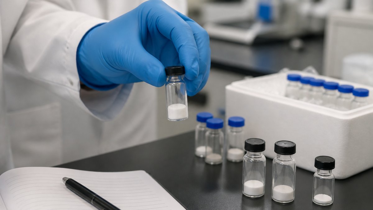
If you buy research grade metabolic peptides for a GLP-1 or incretin study, the documentation that ships with the vial matters as much as the compound. Bluegrass Research supplies research-grade metabolic peptides with lot-specific, third-party analytical documentation, because a certificate of analysis is the only defensible record of what is in the vial. Without it, your experimental design rests on an unverified assumption.
Metabolic peptides are synthetic compounds designed to interact with specific biological targets: GLP-1 receptors, incretin pathways, and the metabolic biology governing glucose handling and energy balance. A supplier's label tells you what the compound is supposed to be; a COA tells you what the lot actually contains.
That distinction shapes everything below: how to read analytical documentation, verify purity certificates before ordering, store lyophilized material, and understand GLP-1 and incretin signaling at the receptor level.
Third-Party Analytical Documentation for Peptides: What to Expect
Third-party analytical documentation for peptides is a lot-specific report from a laboratory independent of the manufacturer, covering identity, purity, and often mass confirmation. A credible package includes an HPLC purity trace, a mass spectrometry result confirming molecular weight, and the analytical methods used.
Here is what a complete documentation set typically contains:
- HPLC analysis with the purity trace and the percentage of the main peak
- Mass spectrometry data confirming the observed molecular weight against the theoretical value
- Amino acid sequence confirmation for the specific peptide
- Lot number that matches the vial label exactly
- Testing laboratory name and the date of analysis
What most buyers miss is that a COA can be technically real and still useless. A report with no lot number, no method description, or a testing date years before production tells you nothing about the vial in your hand. The document must be traceable to your specific lot.
Reading a Certificate of Analysis: Key Fields and Red Flags
Read a COA by checking four fields in order: lot number, purity result, analytical method, and testing date. If any is missing or inconsistent with the vial, treat the documentation as incomplete.
Red flags worth naming directly:
- A purity figure with no accompanying chromatogram
- A lot number on the COA that does not match the vial label
- Mass spectrometry results reported without the theoretical molecular weight for comparison
- The same COA reused across multiple lots
- No named testing laboratory
The FDA guidance on analytical procedures and methods validation sets out the general expectation that analytical methods be documented and validated, which is the standard your supplier's paperwork should reflect.
How to Verify Peptide Purity Certificates Before You Order
Verification starts before payment. Ask for the lot-specific COA for the exact lot being shipped, then confirm the testing laboratory is independent of the manufacturer.
A short checklist you can run on any supplier:
- Request the COA for the specific lot, not a sample document
- Confirm the lot number appears on both the COA and the vial label
- Check that the testing laboratory is named and third-party
- Verify the analytical method is stated (HPLC, mass spectrometry)
- Confirm the analysis date is recent relative to the production date
- Ask whether the COA is accessible online for the lot you receive
Cross-Referencing Lot Numbers and Analytical Methods
Cross-reference the lot number across three places: the vial label, the COA, and the supplier's online record. If a supplier cannot show a lot number before shipping, you cannot confirm the material was ever tested.
The analytical method matters just as much. HPLC separates compounds by retention time and reports purity as a percentage of the main peak. Mass spectrometry confirms the observed molecular weight matches the theoretical value for your peptide's amino acid sequence. Together they confirm identity and purity; either alone leaves a gap.
Best Practices for Peptide Storage in Laboratories
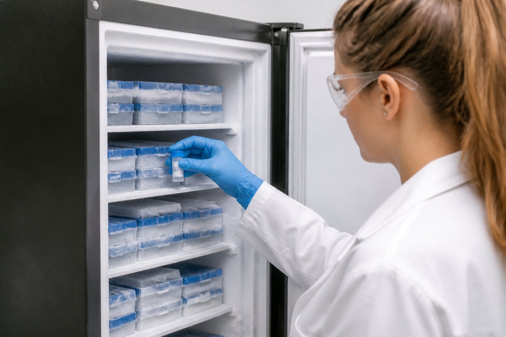
Storage decisions determine whether the purity documented on your COA still describes the material six months later. The rules below hold up in practice, with the reasoning behind each.
Lyophilized Storage
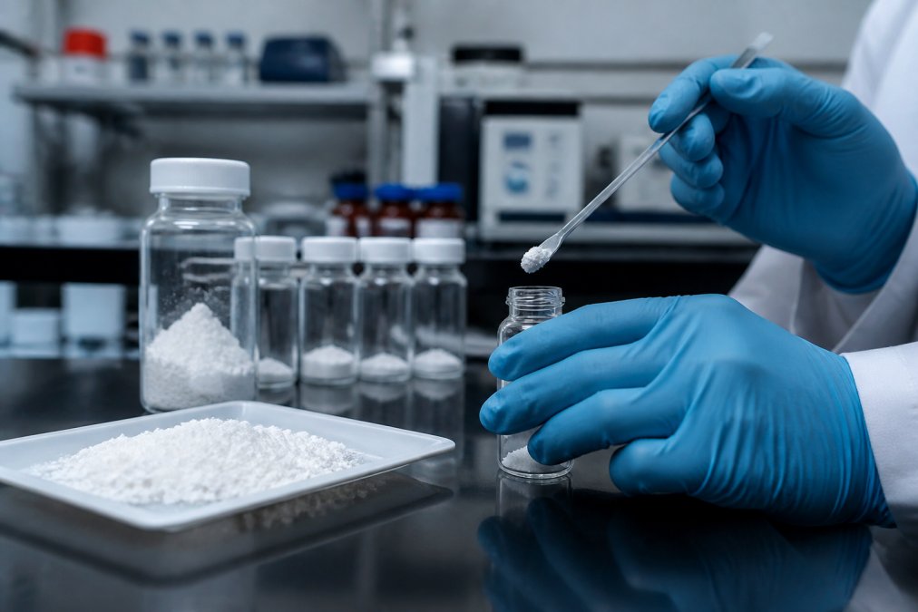
Lyophilized (freeze-dried) peptide is the most stable form. Store it as follows:
- Temperature. Long-term storage is typically at -20 degrees Celsius or colder; -80 degrees Celsius is common for peptides intended to be held for more than a year (peer-reviewed research). A frost-free freezer cycles above and below its set point, so a manual-defrost or laboratory-grade unit is preferred.
- Light. Amber vials or opaque secondary packaging protect against photodegradation, which is a particular concern for peptides containing tryptophan, tyrosine, or methionine.
- Moisture. Keep the desiccant in the vial cap in place and do not open vials until they have equilibrated to room temperature. Opening a cold vial in a warm, humid room pulls condensation directly onto the powder.
- Headspace. Minimize the number of times a vial is opened. Each opening exchanges the dry headspace for room air.
Reconstitution
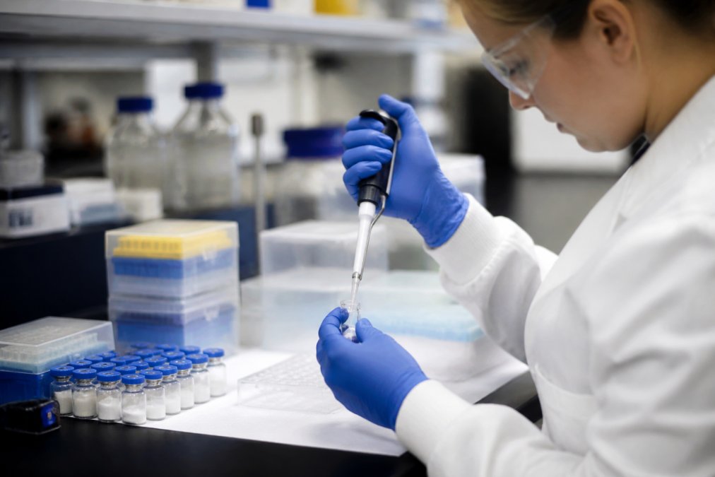
Reconstitution is where most material is lost. Key steps:
- Equilibrate. Let the sealed vial sit at room temperature for 15 to 30 minutes before opening.
- Choose the solvent. Sterile water for most peptides; dilute acetic acid (commonly 0.1 percent) for peptides that are poorly soluble in water; dilute ammonium hydroxide for the most basic sequences. Bacteriostatic water containing 0.9 percent benzyl alcohol is convenient for multi-draw vials but the preservative can interfere with some cell-based assays (peer-reviewed research).
- Add solvent down the wall. Direct the stream at the glass, not at the powder, to avoid foaming and shear.
- Swirl, do not vortex. Vortexing generates shear forces that can denature or aggregate peptide. Gentle swirling or inversion is sufficient.
- Check clarity. A properly dissolved solution is clear. Persistent cloudiness suggests incomplete dissolution, aggregation, or a solubility problem with the sequence.
Concentration Math
Reconstitution concentration is set by the mass of peptide in the vial and the volume of solvent added. A 5 mg vial plus 1 mL of solvent yields a 5 mg/mL stock (5,000 micrograms per mL); a 100 micrograms per mL working concentration is a 1:50 dilution. Record the stock concentration on the vial label at reconstitution, reconstructing it later from memory is a common source of error.
Storage of Reconstituted Solution
- Aliquot before freezing. Divide the reconstituted solution into single-use aliquots sized to one experiment. This is the single most effective step against freeze-thaw damage.
- Freeze at -20 degrees Celsius or colder. Reconstituted peptide is less stable than lyophilized powder; most sequences are usable for a few weeks refrigerated or a few months frozen, but the exact window is sequence-dependent.
- Avoid repeated freeze-thaw. Each cycle promotes aggregation and, for some sequences, deamidation. If you must thaw, thaw on ice and use the aliquot fully.
- Protect from light. Wrap aliquots in foil or store in an opaque box.
A Standardized Protocol Template
A written protocol that travels with the material prevents the most common reproducibility failures. At minimum, record:
- Lot number and supplier
- Date of receipt and date of reconstitution
- Solvent used and final stock concentration
- Aliquot volume and storage temperature
- Date each aliquot was thawed and used
A common mistake is reconstituting an entire vial and refreezing it repeatedly. Each cycle costs material integrity. Aliquot first, then freeze.
Mechanism of Action: GLP-1, Incretin Signaling, and Metabolic Pathways
GLP-1 is an incretin hormone released from intestinal L-cells after a meal. It binds the GLP-1 receptor (GLP-1R), a class B G-protein-coupled receptor expressed on pancreatic beta cells, subsets of neurons, and several peripheral tissues. That binding begins the signaling cascade research-grade metabolic peptides are designed to probe.
The cascade runs in a predictable order, and understanding it separates a useful study from a vial on a shelf:
- Ligand binding. The peptide docks into the extracellular domain of GLP-1R. Affinity at this step depends on the intact amino acid sequence and on the secondary structure of the peptide.
- G-protein activation. The occupied receptor acts as a guanine nucleotide exchange factor for the Gs alpha subunit, which exchanges GDP for GTP.
- Adenylyl cyclase activation. Activated Gs alpha stimulates adenylyl cyclase, which converts ATP to cyclic AMP (cAMP).
- Downstream effectors. Rising cAMP activates protein kinase A (PKA) and the exchange protein directly activated by cAMP (EPAC). In beta cells, these effectors drive insulin granule exocytosis in a glucose-dependent manner, insulin release is amplified only when blood glucose is already elevated.
- Receptor desensitization. After sustained stimulation, GLP-1R is phosphorylated by G-protein-coupled receptor kinases and recruited to beta-arrestin, which uncouples it from Gs and promotes internalization. This is why chronic exposure and acute exposure can produce different readouts in the same assay.
Two features of this pathway matter for experimental design. First, the glucose dependence of insulin secretion means a GLP-1 analog can look inactive in a low-glucose assay even when the compound is fully potent. Second, cAMP is a transient second messenger; the time point at which you sample it changes the number you get.
Why Purity and Potency Are Not the Same Measurement
A peptide can pass HPLC at 98 percent purity and still underperform functionally. Common reasons:
- Deamidation of asparagine or glutamine residues, which shifts mass by roughly one dalton and can blunt receptor affinity without changing the main HPLC peak much.
- Oxidation of methionine or tryptophan residues, which is accelerated by dissolved oxygen and by repeated freeze-thaw cycles.
- Aggregation, which reduces the effective concentration of monomeric, receptor-competent peptide.
- Truncation during synthesis, which produces a shorter sequence that may still co-elute with the full-length product on some HPLC methods.
This is why a mass spectrometry result confirming observed molecular weight against the theoretical value is not redundant with an HPLC purity figure. HPLC tells you how much of the main peak is present; mass spectrometry tells you whether that peak is the right molecule.
Assay Considerations for Incretin Research
When you design a study around GLP-1R signaling, three variables drive most of the variability:
- Glucose concentration in the buffer. Because insulin secretion is glucose-dependent, a low-glucose condition can mask a potent compound.
- cAMP assay format. Competitive immunoassays and FRET-based biosensors have different dynamic ranges and different sensitivity to phosphodiesterase activity. Adding a phosphodiesterase inhibitor changes the readout.
- Incubation time. Acute (minutes) and chronic (hours to days) exposures engage different arms of the pathway because of beta-arrestin-mediated desensitization.
For your experimental design, this means two things: confirm identity and purity through HPLC and mass spectrometry before trusting a potency assumption, and control storage and handling, because receptor binding and biological activity depend on the compound arriving intact.
Safety, Regulatory Compliance, and Batch Consistency for Research Use
Research-grade peptides are sold for laboratory use only and are not drugs, supplements, or products for human or veterinary use. Suppliers operating within the United States should be clear about that distinction, and researchers should keep institutional compliance requirements in view when sourcing.
Batch-to-batch consistency is the quiet variable in long studies. If lots differ in purity or in the analytical method used to test them, results may not be comparable across phases. Ask how a supplier controls consistency: whether each lot is tested independently, whether the same methods are applied, and whether historical COAs are retained.
Institutional purchasing adds a practical wrinkle. University and biotech procurement teams often require documentation matching their compliance checks, so confirm early that the supplier's paperwork and shipping fit your institution's process. Discreet shipping with no external branding is useful, but the documentation still has to satisfy procurement.
Conclusion
Sourcing research-grade metabolic peptides is a documentation problem before it is a purchasing problem. The suppliers worth your budget can show a lot-specific COA, name an independent testing laboratory, and keep analytical methods consistent from lot to lot. Everything else, purity claims, potency claims, even packaging, depends on that foundation.
Bluegrass Research was built around that requirement. Every compound ships with lot-specific COA documentation and third-party analytical testing, so purity and identity are verifiable rather than assumed. Discreet shipping with no external branding keeps your supply chain quiet, and documentation is treated as part of the product, not an afterthought.
Get started with Bluegrass Research and give your next study the verified documentation it needs to hold up under scrutiny.
Frequently Asked Questions
What documentation should accompany research grade metabolic peptides?
Every order of research grade metabolic peptides should include a lot-specific Certificate of Analysis (COA) with HPLC purity results, mass spectrometry data confirming molecular weight, and the amino acid sequence. Look for third-party analytical documentation for peptides that lists the testing laboratory, method parameters, and lot number matching your vial. A COA without a lot number or testing date is not sufficient for rigorous laboratory use.
How do you verify the purity of peptides for laboratory research?
Start by checking the COA for HPLC purity percentage and the chromatogram itself. Cross-reference the lot number on the COA with the vial label. Confirm that mass spectrometry data matches the expected molecular weight for your specific peptide. For critical experiments, consider independent testing through an accredited lab. Reputable suppliers will provide full analytical documentation and respond to requests for additional verification data.
How should research grade peptides be stored to maintain integrity?
Lyophilized peptides should be stored at -20°C or lower, protected from light and moisture. Once reconstituted, aliquot into single-use portions to avoid repeated freeze-thaw cycles, which degrade peptide stability. Use sterile technique and appropriate buffers. Keep a storage log documenting temperature, date, and handling. For long-term storage, -80°C is preferred for most metabolic peptides.
What are the risks of sourcing low-quality metabolic peptides?
Low-quality peptides may have incorrect amino acid sequences, low purity with unknown contaminants, or degraded material from improper shipping and storage. These issues can invalidate experimental results, waste research budgets, and potentially introduce safety concerns. Without third-party analytical documentation for peptides, you cannot confirm identity or purity. Always request lot-specific COAs and verify the supplier's quality control processes before purchasing.